鼻中隔
外观
| 鼻中隔 | |
|---|---|
 Bones and cartilages of septum of nose. Right side. | |
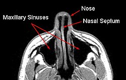
 MRI image showing nasal septum. | |
| 基本信息 | |
| 动脉 | Anterior ethmoidal posterior ethmoidal sphenopalatine greater palatine branch of superior labial[1] |
| 神经 | Anterior ethmoidal nasopalatine nerves[1] Medial posterosuperior nasal branches of pterygopalatine ganglion |
| 淋巴 | Anterior half to submandibular nodes Posterior half to retropharyngeal and deep cervical lymph nodes |
| 标识字符 | |
| 拉丁文 | Septum nasi |
| MeSH | D009300 |
| TA98 | A06.1.02.004 |
| TA2 | 3137 |
| FMA | FMA:54375 |
| 格雷氏 | p.993 |
| 《解剖學術語》 [在维基数据上编辑] | |
鼻中隔(Nasal septum)把人类鼻内分成两半,形成两个鼻孔。鼻中隔由筛骨,犁骨和四方形的软骨组成。
鼻中隔一般为处在鼻内中央位置,但由于天生或者后天外伤可能导致鼻中隔偏曲,堵塞空气正常经过鼻孔,一般不影响生活,但严重时可导致用鼻呼吸困难,经常流鼻血,和反复炎症,需用手术治疗。
附加圖片
[编辑]-
Horizontal section of nasal and orbital cavities.
-
Left orbicularis oculi, seen from behind.
-
Cartilages of the nose, seen from below.
-
Nasal septum
-
Coronal section of nasal cavities.
-
Front of nasal part of pharynx, as seen with the laryngoscope.
-
MRI image showing nasal septum.
-
MRI image showing deviated septum
註釋
[编辑]外部連接
[编辑]- Anatomy figure: 33:02-01 at Human Anatomy Online, SUNY Downstate Medical Center - "Diagram of skeleton of medial (septal) nasal wall."
- Diagram at evmsent.org
- (英文)lesson9 在韦斯利诺曼的解剖课上(乔治城大学) (nasalseptumbonescarti)
| 这是一篇與解剖學相關的小作品。您可以通过编辑或修订扩充其内容。 |


